Sound Field Simulation in Breast Cancer Research
Dinah Brandner

According to the American Cancer Society, it is estimated that one in eight women will develop invasive breast cancer in their lifetime. Key for a full recovery is early detection, which is why regular screening is recommended, as many early breast carcinoma show no symptoms like pain or discomfort, and are too small to be detected by self-observation. The gold standard for breast screening still is the mammography, being the most accurate in diagnosis, besides magnetic resonance imaging (MRI) and ultrasonic imaging. However, since screening by mammography or MRI is associated with risks and comparatively high costs, efforts are being made to increase the diagnostic accuracy of the noninvasive ultrasound imaging.
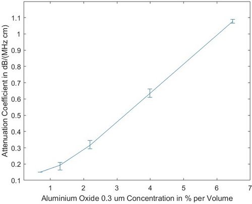
This can be done by estimation of the acoustic properties of the different breast tissue types out of ultrasound image data, as being able to determine these properties additionally to the structural (morphologic) information, is expected to improve the diagnostic accuracy. But, in order to verify that the method used for estimation leads to unbiased results of low variance, methods for producing phantoms of known locally variable acoustic properties need to be developed. Part of this thesis is therefore devoted to the improvements of methods for the production of phantoms with certain acoustic properties, using tissue-mimicking materials. Since it is known from literature that the majority of malignant processes are highly acoustically attenuating, formulas based on Al2O3 are characterized with respect to their absorption behavior, as this material, dependent on the chosen concentration, allows to select the attenuation parameters of a to be produced phantom (see Figure 1).
 Figure 1: Attenuation coefficient vs. concentration of aluminium oxide 0.3 μm for
homogeneous tissue-mimicking phantoms.
Also artificial intelligence can help in the improvement of ultrasonic diagnostic, when employed to assist the radiologist in making a diagnosis by drawing attention to even small morphologies that may seem suspicious. Using huge annotated databases and self-learning algorithms, computers can recognize trained patterns with great statistical certainty. These annotated databases are not yet available for ultrasound images or not in sufficient quality.
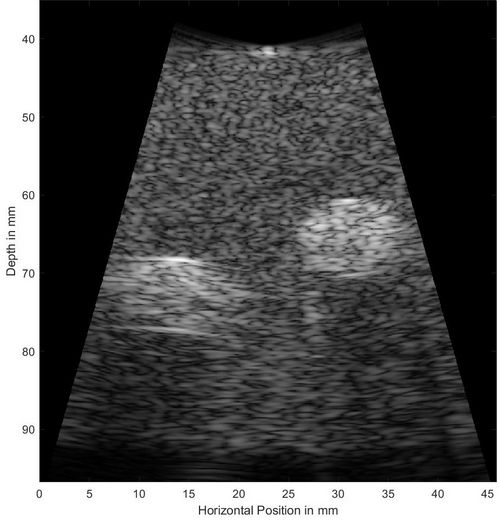
Therefore, ultrasound simulations of annotated volume data derived from other imaging modalities are useful for the generation of a breast cancer ultrasound database. Two open-source ultrasound simulation software, Field II, and k-Wave, were tested for suitability of providing valid simulation results that match as closely as possible in any aspect ultrasound images obtained from clinical equipment. As it turned out k-Wave was designed to fullfill all necessary requirements, and was therefore investigated in great detail. An example of a simulation as presented in the thesis is depicted in Figure 2.
 Figure 2: Simulated B-mode image of a phantom with a low attenuating background
(α_0 = 0:3 dB/(MHz cm)), and two highly scattering regions of α_0 = 1.2, and 2.3
dB/(MHz cm), respectively.
Keywords: ultrasound-simulations, k-Wave, tissue-mimicking material, breastcancer, deep learning
July 14th, 2020
 Go to JKU Homepage


