 Go to JKU Homepage

Dipl.-Ing. Dr. Edgar Scherleitner

Supervisory committee: | |
|---|---|
Univ.-Prof. Dipl.-Ing. Dr. Bernhard Zagar | |
Final exam: | |
November 24, 2005 | |
Supervisory committee: | Univ.-Prof. Dipl.-Ing. Dr. Bernhard Zagar |
Final exam: | November 24, 2005 |
In the presented work a method of optical tomography is introduced which is suitable for investigating diffuse objects, for example soft human tissues, to detect inhomogeneities having deviating absorption- and scattering properties, respectively, compared to the surrounding volume. Intensity modulated near infrared light, is coupled into the surface of the object and is picked up amplitude damped and phase shifted at a different surface location. The set of amplitude ratios and phase differences obtained by various such geometrically distributed measurements create a record which becomes a part of a system of equations to be approximately solved by inversion. A scan-head, which guides the attached optical source- and detector fiber over the surface, was developed to easily collect the desired set of records. The number and the geometrical distribution of the pickup locations was optimized for the setup and various investigated objects. Eventually a mathematical method was developed to obtain slice images to localize potential inclusions by justifiable effort, this turned out to be possible for certain geometries and optical properties of tested probes. The first part explains in detail the theory of light propagation in turbid media and methods are evolved to obtain utilizable information of the inner structure of a probe. Finally finite elements simulations of probes containing various inclusions are performed as well as real measurements of similar probes to be compared with each other. The work concludes by discussing the applicability of this system in real-life measurements and arising problems.
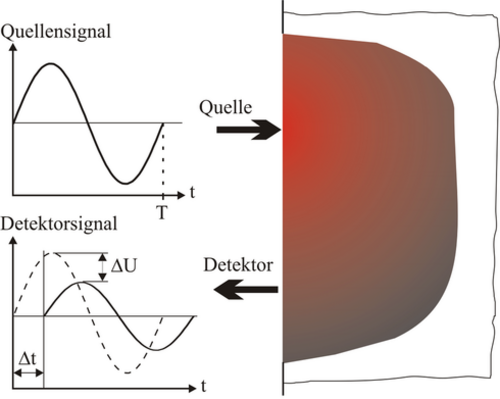 Figure 1: Intensity modulated light is used to determine an amplitude difference and a time delay between the source and the detector signal. Optical inhomogeneities in a mostly homogenous medium can be located by a number of such measurements at different positions on the surface.
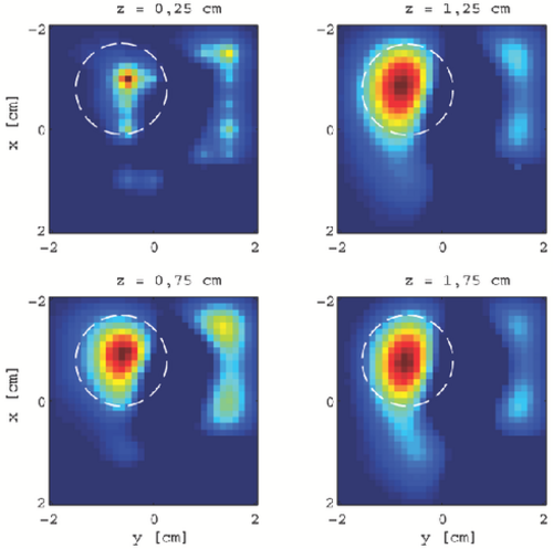 Figure 2: This figure shows four slices of an examined forearm area with a lipoma. The real position and size of the lipoma was fumbled and marked.
