Since 2021, the JKU medSPACE has been delivering fascinating educational insight into the human body.

New, innovative, and impressive data sets generated by the synchrotron in Grenoble support advancements in medical education.
The large-scale, multinational research facility European Synchrotron Radiation Facility (ESRF) in Grenoble (France) operates the largest electron synchrotron in Europe, the third largest of its kind in the world. The synchrotron in Grenoble uses an enormously powerful radiation source to conduct research and examine human donated organ samples.
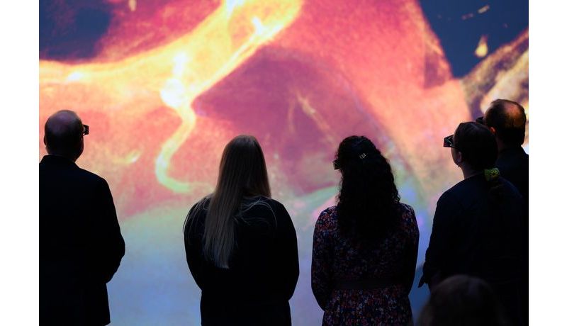
The JKU medSPACE recently installed a number of these data sets to display stereoscopic 3D images onto a 14x7 m projection screen during university lectures and for other presentations. The data set resolution is less than 50 micrometers, displaying high-resolution anatomical structures barely visible to the naked eye but more impressively, those that are invisible to the naked eye.
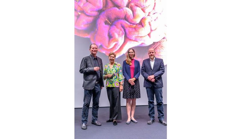
Elgin Drda, Vice-Rector at the JKU’s Faculty of Medicine, remarked: "Understanding anatomy is essential in order to successfully study medicine. The JKU medSPACE is unique, taking pre-clinical education at our Faculty of Medicine to a whole new level. The new data sets from Grenoble provide an even more impressive 3D journey into the human body. I am delighted that we here at the JKU medSPACE are able to not only inspire our students, but also anyone else interested in the fascinating world of anatomy through our free 'Anatomy for Everyone’ series."
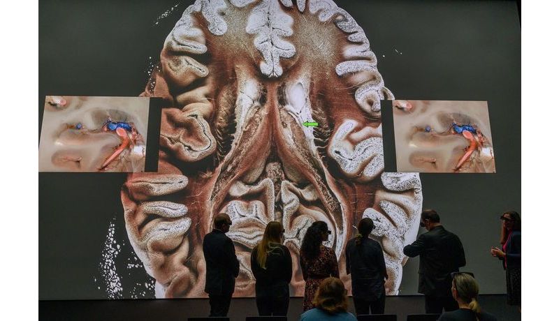
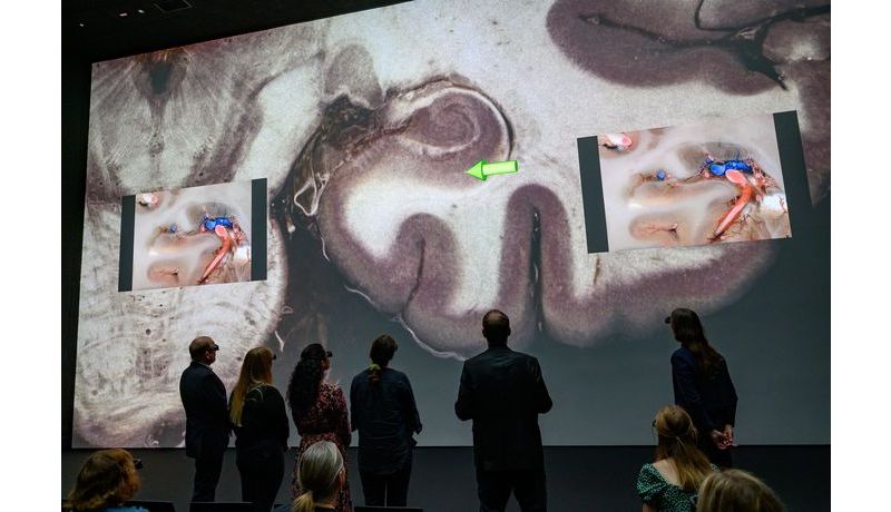
The new data sets are unique, allowing the user to view a specific organ and continuously zoom back and forth between the macroscopic and microscopic range. When it comes to teaching anatomy and supporting post-graduate studies, the new data sets offer new options, particularly to study the brain, heart, and kidneys in more detail. The synchrotron can generate cross-sectional images, making it possible to zoom in to an overall organ image - such as the brain, for example - to a detailed microscopic image from inside of the brain, thereby revealing specific details, structures, fibers, and neural connections.
Professor Franz Fellner prepares the new data sets specifically for university lectures and presentations, projecting the ultra-high, 3D resolution sets during virtual anatomy and pathoanatomy courses onto a 14x7-meter size screen at the JKU medSPACE.
Prof. Franz Fellner enthusiastically added: "The synchrotron’s data sets give our med students an even deeper understanding of anatomy and pathology. The same applies to our public-accessible anatomy presentations, as well as the interdisciplinary educational courses for medical professionals. These data sets have further improved the quality of these courses."
Klaus Engel, Senior Principal Key Expert at Siemens Healthineers, prepared the data sets and a team from the Ars Electronica Future Lab installed the sets at the JKU medSPACE. Engel remarked: "Physicists often talk about an extremely brilliant radiation source, the EBS (Extremely Brilliant Source), but in fact, the resolution currently ranges at around 25 micrometers for the entire organ, to close to micron resolution in partial areas. Researchers soon hope to record data sets in nanometer resolution. The huge amount of data - which will soon be several terabytes per organ – is very challenging in terms of analysis and rendering."
Prof. Maren Engelhardt commented: "Unlike at other Austrian medical universities, the JKU has integrated histology and macroscopic anatomy into one of the three basic areas within the study of anatomy. Our students can really get 'up close and personal' with the cellular structure of human organs. Bringing histology into the JKU medSPACE is an enormous challenge but the new data sets are helping us make important strides."